
Tocotrienols Analyzed by HPLC - AppNote
August 5, 2026
/
/
/
/
Date: 05-August-2026 Last Updated:
06-August-2026
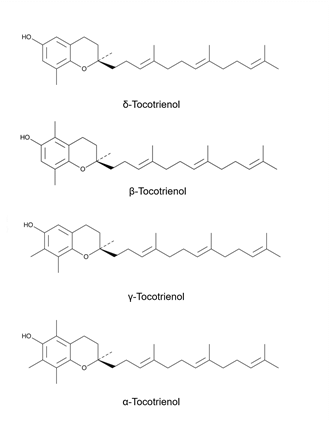
Tocotrienols can present a significant chromatographic challenge due to their closely related structures and physicochemical properties. The α-, β-, γ-, and δ-forms differ only in the number and position of methyl groups on the chromanol ring, resulting in very similar chromatographic behavior and often leading to co-elution or poor resolution with conventional HPLC methods.aration essential for research and quality control.
The ability to achieve separation of all all four tocotrienols provides a powerful tool for vitamin E profiling in nutraceuticals, edible oils, and dietary supplements.
Method Conditions:
Column: Cogent Bidentate C18™, 4 μm, 100 Å
Catalog No.: 40018-25P
Dimensions: 4.6 x 250 mm
Mobile Phase: 25% DI Water / 75% Acetonitrile / 0.1% formic acid
Note: Although all four tocotrienols belong to the vitamin E family, they differ only in the number and position of methyl groups on the chromanol ring. These subtle structural differences can significantly affect biological activity, making accurate chromatographic separation essential for research and quality control.
Tocotrienols can present a significant chromatographic challenge due to their closely related structures and physicochemical properties. The α-, β-, γ-, and δ-forms differ only in the number and position of methyl groups on the chromanol ring, resulting in very similar chromatographic behavior and often leading to co-elution or poor resolution with conventional HPLC methods.aration essential for research and quality control.
The ability to achieve separation of all all four tocotrienols provides a powerful tool for vitamin E profiling in nutraceuticals, edible oils, and dietary supplements.
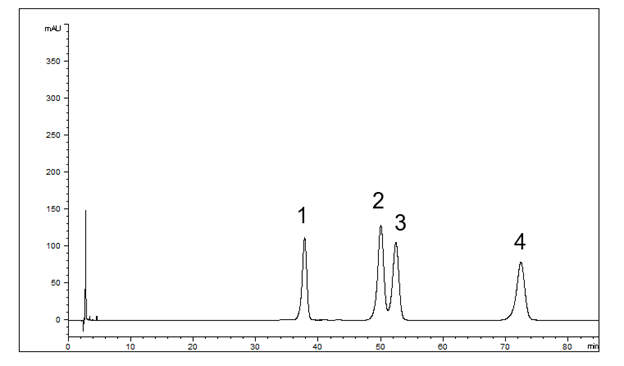
Peaks:
1.
δ-Tocotrienol, 2. β-Tocotrienol, 3. γ-Tocotrienol, 4. α-Tocotrienol
Method Conditions:
Column: Cogent Bidentate C18™, 4 μm, 100 Å
Catalog No.: 40018-25P
Dimensions: 4.6 x 250 mm
Mobile Phase: 25% DI Water / 75% Acetonitrile / 0.1% formic acid
Flow Rate:
1.0 mL / minute
Detection: 220 nm
Injection Volume: 1 uL
Sample Preparation: β-Tocotrienol, α-Tocotrienol, γ-Tocotrienol, δ-Tocotrienol were each prepared as a 1.0 mg / mL concentration in MeOH.
Detection: 220 nm
Injection Volume: 1 uL
Sample Preparation: β-Tocotrienol, α-Tocotrienol, γ-Tocotrienol, δ-Tocotrienol were each prepared as a 1.0 mg / mL concentration in MeOH.
Note: Although all four tocotrienols belong to the vitamin E family, they differ only in the number and position of methyl groups on the chromanol ring. These subtle structural differences can significantly affect biological activity, making accurate chromatographic separation essential for research and quality control.